Mirror Therapy for Relief from Phantom Limb Pain Inspired By Professor Ramachandran

Feature Image of Professor Ramachandran sourced from:

https://www.thehindu.com/news/cities/chennai/Demystifiying-a-phantom/article17009821.ece 

Thanks to Professor Ramachandran’s inspiring research into mirror therapy, the world is now a better place for many amputees with phantom limb pain and other patients!

For related blog posts, see:

https://painmatters.wordpress.com/2019/02/14/mirror-mirror-short-or-tall-which-patient-has-no-pain-at-all

https://painmatters.wordpress.com/2019/02/18/mirror-therapy-what-your-eyes-see-is-reality

https://painmatters.wordpress.com/2019/02/19/a-ghost-foot-and-a-phantom-leg-treated-by-mirror-therapy

Dear Pain Matters blog readers,

This blog post discusses:

  • A clinical trial involving mirror therapy;
  • Two (2) amputees who had successful mirror therapy for phantom limb pain; and
  • Professor V.S. Ramachandran’s ground-breaking research into mirror visual feedback via mirror therapy.

Phantom pain affects at least 90% of all amputees.  It includes a myriad of painful sensations including the following:

  • The limb feels frozen in an awkward position;
  • Burning;
  • Stabbing;
  • Shooting; or
  • Electrical shocks.

The good news is that mirror therapy (aka mirror visual feedback) may offer a cheap and effective treatment option for some amputees with phantom limb pain and other pain sufferers.

WALLACE FANENE, THE EXPLOSION AND THE TRAUMATIC LOSS OF 2 LIMBS

Sgt. Wallace Fanene’s Story  

Sgt. Wallace (‘Wally’) Fanene, a 25-year old Samoan in California, was severely injured on September 8, 2007, while serving in Iraq.  He lost both his right arm and his right leg in an improvised explosive device (IED) blast.  At first, Wally did not feel any pain, although he had a burning sensation in his injured legs.  In his words,

‘I knelt down on a landmine;  I sat on it.  I knew my leg was gone right when I was hit, but I didn’t feel any pain, and I didn’t know about my arm.’

Things got even weirder at the hospital when Wally asked his wife to scratch his bandaged right hand to relieve itching.  What Wally did not realise was that his lower right arm was also missing!

[My wife] told me my arm was gone. We argued about that for five minutes. I mean, I could feel it.’

It was at the hospital when the pain kicked in big time, even affecting his sleep.  There were nails in his phantom toes as well as needles and a knife-like pain in his missing leg.

Wally added,

‘It was the most excruciating pain in my life.’

arm_gone_t300.png

Source: https://www.sandiegoreader.com/news/2008/jul/09/cover/

Enter Mirror Therapy  

Not knowing what to expect, Wally enjoyed immediate pain relief as soon as he started mirror therapy.  

In Wally’s words,

‘I was sceptical at first, but then I said, what the heck. So we tried it and [mirror therapy] worked right away. The pain just stopped. Four hours later the pain came back again, and again the mirror helped right away … 

… The pain still comes back, but it’s nowhere as bad as it was.  It comes back several times a day.  But if [the phantom limb pain] starts to bother me, I use the mirror.’

Wallace+Fanene+San+Onofre+Beach+Host+Surf+pRIAxYjhj9-l.jpg

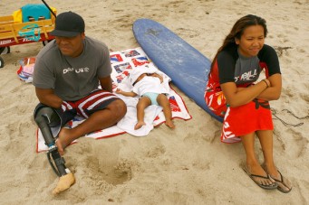
Wallace Fanene, double amputee, and his wife

https://www.gettyimages.co.uk/photos/nalia-fanene?sort=mostpopular&mediatype=photography&phrase=nalia%20fanene

A CLINICAL TRIAL INVOLVING MIRROR THERAPY 

Overview

Inspired by Professor Ramachandran’s ground-breaking research into mirror visual feedback in the mid-1990’s, Jack Tsao, Neurologist, offered mirror therapy for his patients who suffered phantom limb pain.

Soon after, Tsao set up a clinical trial involving mirror therapy for phantom pain at the Uniformed Services University in Maryland.

Specifically, 22 lower limb amputee patients with phantom pain from Walter Reed Army Medical Center (WRAMC) were randomly allocated to 1 of 3 groups.  Four (4) patients were unable to complete this study.

The 1st group (N=6) received mirror therapy.  The remaining 2 groups underwent covered mirror treatment (N=6) and mental visualisation (N=6).

Each group was instructed to do their assigned therapy for 15 minutes daily for 4 weeks.

This study found that ALL 6 amputees in the first group who received mirror therapy enjoyed reduced phantom limb pain after one month!  Some even experienced complete relief from phantom pain after mirror therapy.

In comparison, half of the 2nd group who received covered mirror treatment and 2/3 of the 3rd group who performed mental visualization exercises had increased pain after 1 month.

Nine (9) amputees from the 2nd and 3rd groups were subsequently re-assigned to classical mirror therapy for 4 weeks.  Eight (8) of these 9 amputees (89%) enjoyed less pain thereafter.

The first group performed 4 additional weeks of mirror therapy, totalling 8 weeks.  This resulted in further improvements for all 6 participants.  

Jack Tsao, Neurologist, said that the results ‘astounded me … I didn’t expect [the results] to be that good.’

Mirror therapy is now available at 3 US military amputee centres including WRAMC as well as other locations for amputees and patients with other painful conditions (Chan et al, 2007; Dobyns, 2008).

Nick Paupore, Amputee and Participant in Above Trial

Nick Paupore (32), a participant in the aforementioned trial, enjoyed significant relief from excruciating phantom pain following mirror therapy.

Nick lost his lower right leg in Iraq when a roadside bomb hit his vehicle.  While he felt a burning sensation in the back of his legs, Nick did not feel any pain.  Unfortunately, the blast tore out a chunk of his leg including 15cm to 20cm of an artery that almost caused him to bleed to death.

At the hospital, Nick suffered excruciating phantom pain in his missing leg 5 – 6 times an hour, with each bout of pain lasting up to a minute.  As he screamed and dry-heaved, the searing shock raced from the missing sole in his foot up the back of his phantom calf and into his thigh.

Quoting Nick,

‘I was laying in bed and it just, all of a sudden, it felt like I was getting shocked.  I called the nurse, ’cause I was like, ‘What’s going on?’  

The nurse told Nick that it was likely his phantom pain.

Nick continued,

‘All of a sudden, it was like someone kept turning on and off the Taser, and my whole leg started twitching. … I sat up, and I was holding on to my stump, and it just wouldn’t stop.’

‘It was like sticking my finger in a light socket. … getting electrocuted.’

‘… the [phantom] pain was driving me insane.’

And now for the good news:

Nick was almost pain-free after mirror therapy for a month.  Furthermore, he no longer needed pain medication 5 months later.

Jack Tsao

Jack Tsao, Neurologist, with Nick Paupore during mirror therapy

http://www.dana.org/QA/Mirror_Therapy_for_Phantom_Limb_Pain/#_ftn2

Quoting Tsao, Neurologist,

‘The difference is like night and day … To see him walking, he’s able to drive his car; he works downtown; I mean, that is incredibly gratifying!’ (Burgess, 2008; Young, 2008).

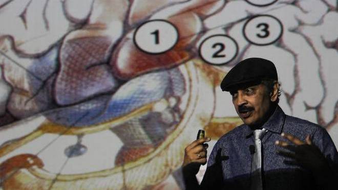
SEMINAL RESEARCH BY PROFESSOR RAMACHANDRAN

Overview

Professor Ramachandran did research in the mid-1990’s based on amputees who had lost their limbs many years earlier.  His studies led to mirror therapy as a pain treatment option for phantom limb pain and other painful conditions.

Ramachandran.jpg

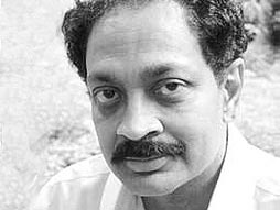
Neuroscientist V.S. Ramachandran

Source: https://greatergood.berkeley.edu/article/item/do_mirror_neurons_give_empathy

Many ground-breaking research findings were reported by Ramachandran including:

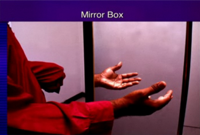
(1) An amputee named ‘D.S.’ lost his left arm 9 years ago.  His phantom arm felt as if it was ‘frozen … in a cement block’.  Ramachandran offered mirror therapy involving bilateral mirror-symmetric movements.  During this therapy, D.S. felt movement in the muscles and joints of his phantom arm while looking at his intact arm in the mirror.

When Ramachandran removed the mirror, D.S. was no longer able to feel his phantom arm move.  In his words, ‘It feels frozen again.’   

This experiment demonstrates the importance of visual feedback via mirror therapy in restoring movement in ‘frozen’ phantom limbs.  This movement may even be a prerequisite before phantom limb pain can be reduced or eliminated altogether.

(2) A left arm amputee known as ‘R.T.’ (55) suffered a paralysed phantom arm.  His left hand often had clenching spasms with ‘fingernails digging into the palm’.

Mirror visual feedback was done to provide an illusion of a restored left arm.  The reflection of his intact right arm in the mirror provided the visual feedback that R.T. desperately needed before he could finally ‘unclench’ and ‘move’ his paralysed phantom arm in a mirror-symmetric manner.

(3) Five patients suffered painful ‘clenching spasms’ in their phantom hand.  Following mirror therapy, 4 of 5 patients were finally able to ‘open’ their phantom hand leading to relief from painful spasms.

Mirror therapy may lead to new connections in the brain.  This may include novel and/or reactivation of pre-existing neural pathways between both cerebral hemispheres (Ramachandran & Hirstein, 1998).

Graded Mirror Therapy vs Traditional Mirror Therapy

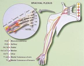
A male patient (43) suffered deafferentation pain in his right arm following brachial plexus avulsion due to a traffic accident 23 years ago.  Specifically, he endured severe tingling pain including numbness in his right fingers (pain rating of 10/10).

Traditional mirror therapy for 4 weeks failed to offer pain relief.

Graded mirror therapy, on the other hand, ultimately led to complete relief from pain.

Graded mirror therapy comprises 5 steps, being:

  1. Observing the mirrored reflection of the unaffected side without imagining any movements in the amputated limb;
  2. Observing the mirrored reflection of the 3rd and 4th fingers while slowly moving them from a flexed position to an extended position;
  3. Observing the mirrored reflection of the unaffected side during passive movements;
  4. Motor imagery of the affected fingers while observing the mirrored reflection of the unaffected side; and
  5. Motor imagery of the affected fingers without a mirror.

Graded mirror therapy involving the aforementioned 5 steps was done for 3 – 4 weeks.

The good news is that during graded mirror therapy, pain levels decreased and finally disappeared altogether.  The patient was also able to imagine finger movement both with and without a mirror (Mibu et al, 2016).

Possible Mechanisms

Although there is no shortage of possible explanations, no one really knows why mirror therapy may work better in some patients than in others.

Professor Herta Flor, Neuroscientist, believes that both peripheral input and central changes may be involved in phantom limb pain (Perur, 2014).

Competitive interaction may arise between the two brain hemispheres in stroke patients (and presumably also in amputees).  This can lead to abnormal interhemispheric inhibition (IHI) and weakened motor function.

In stroke patients, mirror therapy and motor training could increase the excitability of the stroke-affected primary motor cortex (M1).  Specifically, mirror visual feedback training could alter the excitability of the transcallosal pathway and restore IHI back to normal.  This could in turn strengthen motor function in the stroke-affected side.

Thus, mirror visual feedback training may help normalise IHI and restore the hemispheric balance between the stroke-affected (or amputation-affected) M1 and the healthy M1 (Avanzino et al, 2014).

Another study reported that mirror visual feedback via a mirror led to improved motor function in 2 patients who had callosal disconnection.  Based on this, they concluded that mirror therapy that leads to improved motor performance does not affect interhemispheric interaction via corpus callosum (Nojima et al, 2013).  Whether this also occurs in amputees is unknown.

See 5-minute TED-Ed Animations video called ‘The fascinating science of phantom limbs’ by Joshua Pate.

Some Questions to Ponder

Could an amputation lead to abnormal IHI and a hemispheric imbalance between the amputation-affected M1 and the healthy M1?

If yes, could this contribute (in part or in full) to ‘painfully frozen limbs’, ‘paralysed phantom limbs’ and phantom limb pain in amputees?

If so, could mirror visual feedback training using mirrors reduce phantom limb pain and other symptoms in some amputees?

Another Question

In my blog post called:

A Doctor and a Nurse who can Literally Feel Pain in Other People,

we learn that Dr Joel Salinas, a neurologist, has mirror touch synesthesia.  This means that he can literally feel his patients’ pain and tactile sensations.  Automatically triggered by sight, he said that, for example, when he sees someone’s right arm being touched, he also feels a touch on his own left hand – just like in a mirror.

Could Dr Salina’s experience with mirror touch synesthesia offer certain insight into why mirror therapy may offer relief from phantom limb pain in many amputees? 

SUMMARY

Mirror therapy including graded mirror therapy may be a very effective treatment option for amputees with phantom limb pain and other chronic pain sufferers.

Benefits derived from mirror therapy vary from one patient to the next.  According to Professor Christoph Maier, while some amputees are pain-free after only 1 mirror therapy session, others may need to do mirror therapy for the rest of their lives to reduce phantom pain (Liebling, 2010; translated from German).

Who would have thought that a humble mirror could offer relief from excruciating phantom limb pain for some amputees??

Sabina Walker

Blogger, Pain Matters (in WordPress)


REFERENCES

Patient Stories

(1) Dobyns, Stephen. Certain San Diegans feel their missing limbs. San Diego Reader (9 July 2008).

(2) Saundra Young. For amputees, an unlikely painkiller: Mirrors. CNN (19 March 2008).

http://www.cnn.com/2008/HEALTH/03/19/mirror.therapy/index.html

(3) Burgess, Lisa. Mirrors helping amputees fight phantom pain. Stars and Stripes (2/2/2008).

https://www.stripes.com/news/mirrors-helping-amputees-fight-phantom-pain-1.74351

(4) Colapinto, John. Brain Games – The Marco Polo of Neuroscience. The New Yorker (11 May 2009).

https://www.newyorker.com/magazine/2009/05/11/brain-games

Peer-Reviewed Science Papers

(5) Chan BL et al. Mirror therapy for phantom limb pain. N Engl J Med (22 Nov 2007); 357: 2206-2207.

doi: 10.1056/NEJMc071927

https://www.physio-pedia.com/images/1/11/Mirror.NEJM.pdf 

(6) Uniformed Services University of the Health Sciences. Phantom Limb Pain May Be Reduced By Simple Mirror Treatment. Science Daily (24 November 2007).

http://www.sciencedaily.com/releases/2007/11/071123195218.htm

(7) Ramachandran VS & Hirstein W. The perception of phantom limbs. Brain (1998); 121: 1603-30.

http://www.rctn.org/bruno/psc129/handouts/rama2.pdf

(8) Avanzino et al. Training based on mirror visual feedback influences transcallosal communication. Eur J Neurosci (Aug 2014); 40(3): 2581-8.

doi: 10.1111/ejn.12615.

(9) Nojima et al. Mirror visual feedback can induce motor learning in patients with callosal disconnection. Exp Brain Res (May 2013); 227(1): 79-83.

doi: 10.1007/s00221-013-3486-4

(10) Perur, Srinath. The mirror man. mosaic (7 July 2014).

https://mosaicscience.com/story/mirror-man/

(11) Mibu et al. Successful Graded Mirror Therapy in a Patient with Chronic Deafferentation Pain in Whom Traditional Mirror Therapy was Ineffective: A Case Report. Pain Practice (April 2016); 16(4): E62-E69.

https://doi.org/10.1111/papr.12431

https://onlinelibrary.wiley.com/doi/full/10.1111/papr.12431

Non-Fiction Book

(12) Ramachandran VS & S Blakeslee. Phantoms in the Brain – Human Nature and the Architecture of the Mind. HarperCollins Publishers (20 May 1999); pages 1 to 352.

ISBN 9781857028959

(13A) Ramachandran VS. The Tell-Tale Brain – A Neuroscientist’s Quest for What Makes Us Human. W.W. Norton & Co (5 April 2012); pages 1 to 357.

ISBN: 9780099537595

(13B) V.S. Ramachandran’s Tales Of The ‘Tell-Tale Brain’. NPR (14/2/2011).

https://www.npr.org/2011/02/14/133026897/v-s-ramachandrans-tales-of-the-tell-tale-brain

Medical Articles (in German) 

(14) Mit der Spiegeltherapie sind Schmerzen “verlernbar”. Ärzte Zeitung online (22.02.2010).

https://www.aerztezeitung.de/medizin/krankheiten/schmerz/article/590067/spiegeltherapie-schmerzen-verlernbar.html

(15) Liebling, Patricia. Spiegeltherapie hilft gegen Phantomschmerzen. Aachener Zeitung (27 Feb 2010).

https://www.aachener-zeitung.de/ratgeber/gesundheit/spiegeltherapie-hilft-gegen-phantomschmerzen_aid-27029737

(16) Hawlik, Michaela. Leben mit Phantomschmerz. My Handicap (1/2011).

https://www.myhandicap.de/gesundheit/koerperliche-behinderung/amputation-prothese/phantomschmerz-spiegeltherapie/

Other Medical Articles (not discussed in this blog post)

(17) Peterzell, David H. Psychophysical investigations into Ramachandran’s mirror visual feedback for phantom limb pain: video-based variants for unilateral and bilateral amputees, and temporal dynamics of paresthesias. 2016 Society for Imaging Science and Technology (Feb 2016).

doi: 10.2352/ISSN.2470-1173.2016.16HVEI-093

https://www.ingentaconnect.com/contentone/ist/ei/2016/00002016/00000016/art00045?crawler=true

(18) Timms, Jason & Carus, Catherine. Mirror therapy for the alleviation of phantom limb pain following amputation: A literature review. International Journal of Therapy and Rehabilitation (March 2015); 22(3): 135-145.

https://www.magonlinelibrary.com/doi/pdf/10.12968/ijtr.2015.22.3.135

(19) Datta & Dhar. Mirror therapy: An adjunct to conventional pharmacotherapy in phantom limb pain. J Anaesthesia Clin Pharmacol (Oct-Dec 2015); 31(4): 575–578.

https://www.ncbi.nlm.nih.gov/pmc/articles/PMC4676260/

(20A) Ol, Ha Sam et al. Mirror therapy for phantom limb and stump pain: a randomized controlled clinical trial in landmine amputees in Cambodia. Scandinavian Journal of Pain (3 July 2018); 18(4): 603-610.

doi: 10.1515/sjpain-2018-0042

https://www.ncbi.nlm.nih.gov/pubmed/30207289

(20B) Ol, Ha Sam et al. Mirror therapy: Curing phantom pain in landmine victims. Science Discoveries (26.10.2018).

https://sciencediscoveries.degruyter.com/mirror-therapy-curing-phantom-pain-landmine-victims/


A Phantom Leg And Frozen Fingers Treated By Mirror Therapy

‘Disability is only a state of mind.’

Bryan Wagner, Amputee 

Feature Image showing mirror therapy sourced from:

https://brainchemist.wordpress.com/2010/11/25/ted-talk-ramachandran-on-your-mind/

For related blog posts, see:

https://painmatters.wordpress.com/2019/02/21/mirror-therapy-for-relief-from-phantom-limb-pain-inspired-by-professor-ramachandran

https://painmatters.wordpress.com/2019/02/14/mirror-mirror-short-or-tall-which-patient-has-no-pain-at-all

https://painmatters.wordpress.com/2019/02/18/mirror-therapy-what-your-eyes-see-is-reality

INTRODUCTION

Dear Pain Matters blog readers,

Phantom limb pain (PLP; phantom pain) is a terrible affliction that affects up to 90% of all people who have lost a limb, or part thereof, due to trauma, injury, disease or surgery.

Mirror therapy may be an effective and inexpensive pain treatment option for patients with phantom pain, complex regional pain syndrome (CRPS), pain and lost motor function after stroke, hand immobility and pain caused by spinal stenosis and other conditions.

Mirror therapy is sometimes called mirror visual feedback or graded motor imagery.

MIRROR THERAPY FOR PHANTOM LIMB PAIN

Bryan Wagner, Amputee and War Veteran  

Bryan Wagner (23) lost his lower right leg in an improvised explosive device (IED) explosion on 17 December 2007 while serving in Baghdad.  Thereafter, Bryan suffered phantom pain in his missing toes and in the arch of his heel.  The pain included knife-like stabbing pain between his toes and poker-like pain.

Mirror therapy is offered to patients just like Bryan at the Walter Reed Army Medical Centre (WRAMC).  This therapy is done 15 minutes a day, 5 days a week, for 8 consecutive weeks.  If necessary, this cycle is repeated.

During mirror therapy, Bryan stated:

‘It … really feels like you are moving your [missing] foot … It feels like your foot is there …’

If you look very closely at the video below, you will see Bryan’s stump actively participate in all the foot exercises at the same time as the good leg.

It is as if the residual stump is doing all the exercises that the missing foot would be doing, if it were to still exist.  Thus, the stump is acting in lieu of the missing foot!  I don’t know about you but I find this very fascinating!

In other words, the leg stump (that is 100% hidden from Bryan’s view) is moving in complete harmony and unison with the foot’s reflection.  Could it be that optimal results from mirror therapy are possible when this occurs?  (Go to 1:28 minutes.)

Mirror therapy enables the brain to finally ‘feel movement’ again in the (phantom) limb.  Using mirror therapy to restore movement in a missing limb (and in a stump) may be key to reducing phantom limb pain.

Treating Phantom Limb Pain with Mirror Therapy 

Above, a 2-minute YouTube video and narrative by UPIVideo dated 27 July 2009.  NB The narrative may only be available if you open the video in a separate window.


MIRROR THERAPY FOR FROZEN FINGERS IN RIGHT HAND CAUSED BY SPINAL STENOSIS

Bob McKeefery, a long-time fisherman, has spinal stenosis (i.e. tightening or narrowing) of the spinal canal.  Spinal stenosis can lead to compression and ‘pinching’ of the spinal cord and nerve roots.  This can cause pain, cramping, weakness and numbness as well as abnormal signalling between the brain and the body.

Bob suffered ongoing pain and limited mobility in his right hand following several surgeries over a year ago.

In his words,

‘I could move my right arm, but I couldn’t move my right hand.  So 3 of my digits, the last 3, were like ‘frozen shut’ and 2 were frozen open.’

The good news is that mirror therapy led to immediate benefits including complete pain relief for Bob!  

Quoting Bob,

‘All my pain, a year’s worth of pain, went away in one day!’

After only 1 week of mirror therapy, Bob was finally able to use muscles and do movements that he had long given up on.

Bob said,

‘It was impossible last week for me to do this.  I could not ‘palm up’. …’ 

As Bob continued with mirror therapy, 10 minutes each session, several times a day, he has one goal that keeps him going.

Quoting Bob,

‘[My goal] would be to pick up my grandchildren … because I’m not allowed during this crisis … and they are 1 and 3.’

Thanks to mirror therapy, Bob is now one step closer to his goal!

For more details, watch Mirror Therapy Helps Patients Reduce Pain, Gain Mobility in Limbs, by NJTV News (30 April 2015).

IN CLOSING

I hope that these stories will inspire others to try mirror therapy for their phantom pain and other painful conditions.  After all, there is no downside in trying mirror therapy.

Sabina Walker

Blogger, Pain Matters (in WordPress)

painmatters.wordpress.com


KEY

* Tibia bone – One of 2 bones in the lower leg.


Mirror Therapy For Phantom Limb Pain – ‘What Your Eyes See IS Reality’

‘What Your Eyes See Is Reality’

Erez Avramov, Life Rebuilder

Feature Image of Erez Avramov and the mirror box was sourced as a still frame from the YouTube video dated 8 July 2013 (below).

For related blog posts, see:

https://painmatters.wordpress.com/2019/02/21/mirror-therapy-for-relief-from-phantom-limb-pain-inspired-by-professor-ramachandran

https://painmatters.wordpress.com/2019/02/14/mirror-mirror-short-or-tall-which-patient-has-no-pain-at-all

https://painmatters.wordpress.com/2019/02/19/a-ghost-foot-and-a-phantom-leg-treated-by-mirror-therapy

INTRODUCTION

Dear Pain Matters blog readers,

Phantom limb pain (PLP; phantom pain) is a terrible affliction that affects up to 90% of all people who have lost a limb, or part thereof, due to trauma, injury, disease or surgery.

Mirror therapy may be an effective and inexpensive pain treatment option for some patients with phantom pain, complex regional pain syndrome (CRPS), pain after stroke and other pain conditions.

EREZ, AMPUTEE AND LIFE REBUILDER

Erez’s Traumatic Life Events 

Erez Avramov, Life Rebuilder, survived 3 serious motor vehicle accidents (MVA) and a serious heart attack.  Sounding more like an action thriller story than an enriched life story, Erez is a perfect example of ‘what doesn’t kill you, makes you stronger’.

About_Erez_140x65.jpg

http://erezavramov.com/about-erez/

During his first accident, Erez was involved in a serious head-on crash with a semi-truck after his vehicle hit black ice and spun out of control near Merritt, BC, Canada, in November 2010.  He suffered many injuries including fractures to the sternum, hand and ankle, a crushed rib cage as well as a leg that was broken in 17 different places.

Several years and multiple surgeries later, Erez opted for elective amputation to improve mobility of his MVA-injured right leg below the knee (ADV Pulse, 2014)

Erez holds the honour of being the first amputee ever to enter the Dakar Rally race in South America.  Sadly, he had his second near-fatal accident when he crashed his motorbike during one of these desert races.

More recently, Erez had another vehicle accident in December 2017 that was followed by a serious heart attack just one (1) month later.

Being optimistic by nature, Erez always tries to gain valuable insight even from the worst moments in his life.  For his inspirational views, please visit his website:

http://erezavramov.com/about-erez/

Erez’s Mirror Therapy for Phantom Limb Pain  

Following his elective lower right leg amputation, Erez suffered phantom pain.

In his words,

‘… the results and the process [of mirror therapy are] just amazing.’

Erez used a custom-made 3-sided mirror box that included a white-framed mirror.  Hinged on both sides, the mirror box can be unfolded and assembled like a ‘triangular tunnel’.  A plain mirror may also be used.

Erez’s good leg was placed right next to the mirror while his amputated right leg remained well hidden inside this triangular tunnel.

As Erez moved his toes very slowly, he tried to imagine moving the same muscles in his phantom toes at the same time.

Given that he spoke about mirror therapy with such amazing clarity, I will quote Erez from here on (see his YouTube, below),

‘… This is the important part … you move your toes and you try with your mind to move … your missing toes – but when you look at them in the mirror, it’s as if they are there … and you feel your muscles in your residual limb …  Move those toes as if they are there.  

Then I will start to do some rotational work , and I will move both legs in the same direction as if everything is there.  

Because what happens is:  Your brain sees this leg and your foot.  It thinks it is there.

This is the most bizarre thing there is.  It is as if you still have it … You  really have to be open-minded and to accept the fact that what your eyes see is reality … It’s almost like you bypass this reasoning side of your brain …

I will move my muscles.  I will move my residual limb as if I have an intact full foot …

The results were amazing … [inserted: my painful ‘tiger-clawed’ phantom toes were] released, so the pain is gone …

… the protocol is 4 weeks of 10 minutes every day.  You do this for 4 weeks …

… You cover your residual limb.  You use … a … magazine cover, or whatever, … to hide your good foot so you don’t see it, and all you want to see is your reflection of your foot, as if you still have [a] missing limb …’ 

Watch Erez Avramov’s helpful video, here:

‘Mirror Therapy – Personal Success Story’

An 8-minute YouTube dated 8 July 2013

IN CLOSING

Thank you very, very much, Erez, for sharing your powerful story about your personal experience with mirror therapy!

I am sure that your story will inspire others to try mirror therapy for their phantom pain.

Sabina Walker

Blogger, Pain Matters (in WordPress)


REFERENCE

(1) http://erezavramov.com

(2) Amputee Pursues Lifelong Dream to Race the Dakar Rally. ADV Pulse (27/10/14).

http://www.advpulse.com/adv-videos/amputee-pursues-lifelong-dream-to-race-the-dakar-rally/


Mirror, Mirror, Short Or Tall, Which One Has No Pain At All?

Title adapted from:

Mirror, mirror on the wall, who is the fairest of them all?

Feature Image sourced from:

http://langaraprm.com/2013/community/mirror-medicine-a-cure-for-phantom-limb-painstephen-sumner-shares-a-simple-treatment-for-phantom-limb-pain/

For related blog posts, see:

https://painmatters.wordpress.com/2019/02/21/mirror-therapy-for-relief-from-phantom-limb-pain-inspired-by-professor-ramachandran

https://painmatters.wordpress.com/2019/02/18/mirror-therapy-what-your-eyes-see-is-reality

https://painmatters.wordpress.com/2019/02/19/a-ghost-foot-and-a-phantom-leg-treated-by-mirror-therapy

INTRODUCTION

Dear Pain Matters blog readers,

Up to 90% of all amputees suffer from phantom limb pain (PLP), or more simply, phantom pain.

Mirror therapy is an inexpensive pain treatment option for some patients with phantom pain, complex regional pain syndrome (CRPS), pain after stroke and other painful conditions.

Stephen Sumner (aka Mirror Man), an amputee and humanitarian from Vancouver, Canada, was so impressed with the pain-relieving effects of mirror therapy on his phantom pain that he set up his mission called ‘Me and My Mirror’.

STEPHEN SUMNER (‘ME AND MY MIRROR’)  

Reality is merely an illusion, albeit a very persistent one.

Albert Einstein

Stephen’s Story, Before Mirror Therapy

Stephen endured a left above-knee amputation after a truck crashed into his scooter in a hit-and-run accident that left him for dead on a quiet country road near Siena, Italy, one balmy evening in 2004.

Using his words, Stephen ‘suffered terribly … suicidally’ from severe PLP for 4 – 5 years thereafter.  He used to endure ongoing electrifying shocks that shot up his missing left leg and throughout his body.

Quoting Stephen, it was like having ‘lightning bolts through my foot’ in a leg that no longer existed.  His entire body would jolt and spasm uncontrollably, as if he was ‘being spiked with a cattle prod day and night’.  Stephen would be reduced to screaming and tears with no sleep at all during these severely painful bouts.

Stephen added, ‘I could have killed myself.’

Stephen’s phantom toes would be ‘stuck’ in an excruciatingly painful and clenched position.  The pain ‘was like a vice over the back of my heel.  It was like it was being crushed.’  Stephen’s phantom pain felt like ‘crippling electric pulses’ … ‘burning and crushing, but the worst is the itching’.

Stephen stated that his phantom pain was ‘… not in the head, it’s in the limb.’

Stephen’s Story, After Mirror Therapy

Then one day, Stephen read about mirror therapy on-line.  The first time he placed a mirror against his left thigh and looked at the reflection of his right leg (where the left leg used to be), he felt immediate relief.  Five (5) minutes later, his pain was gone.  Stephen did mirror therapy twice daily for 10 minutes at a time.  Finally, after 5 weeks, his phantom pain disappeared for good. 

Quoting Stephen, mirror therapist:

‘… within … 3 and 5 weeks, the pain … disappeared … my phantom pain had gone away, almost magically …’ 

Stephen continued,

‘… 10 minutes per session, 2 sessions per day, 5 weeks, and you’re done for life … If I get some kind of a flare-up, I just whip out the mirror, and I’m good again …’

(Go to the following 10-minute video called The Me & My Mirror Back Story; 2:30 minutes).

Stephen offers mirror therapy to amputees with phantom pain in 3rd world countries including Cambodia, Laos, Vietnam, Sri Lanka, Burma, Myanmar, Vietnam, southern part of India, Lebanon, Ethiopia, Syria, Egypt, Tunisia, Algeria and the western Sahara region.

Stephen often treats amputees with PLP due to traumatically torn or mutilated limbs resulting from war, landmine blasts, unexploded ordnances (cluster bombs, cluster munitions) and road accidents.  Stephen also treats patients who lost their limbs due to severe diabetes and other diseases.

Stephen collaborates with medical experts in the local hospitals, trauma centres, physical rehabilitation centres and prosthetic clinics.  Recently, he was at the ‘Jaffna Teaching Hospital’ with Physical Rehabilitation Therapists and Orthopedic Surgeon.  He also teaches locals how to make therapeutic mirrors for pain relief.

Stephen uses this simple mantra for mirror therapy:

Look

Move

Imagine


cambodia_amputee_assistance_theropy_755.jpg

Source: http://meandmymirror.org

In Stephen’s words,

‘… You feel immediate relief, but you have to carry on for 4 or 5 weeks.  So, my mantra is:

  • 2 sessions a day 
  • 10 minutes per session
  • 5 weeks.’

(Global News, 2016)

DSCF5578.jpg

Source: http://meandmymirror.org

Stephen uses acrylic mirrors due to their cost-effectiveness, safety, lightweight nature and transportability (i.e. on the back of his beloved bicycle).

cambodia_amputee_assistance_theropy_395.jpg

Source: http://meandmymirror.org


According to Stephen, it is often difficult for amputees in Cambodia and other 3rd world countries to open up about their phantom pain.  Many amputees with phantom pain are ashamed.  They would rather suffer in silence than risk being labelled as ‘outright crazy’ or ‘insane’ for ‘complaining’ about pain in a limb that does not even exist.

Also, the fact that many amputees here are Buddhists raises the topic of karma including what the amputees ‘must have done in a previous life to deserve this fate’.

When Stephen is seen riding around on a bicycle with his prosthesis clearly visible to all, the locals are more likely to empathise and connect with him.  When Stephen tells them that he used to suffer from phantom pain, they finally open up about their own phantom pain.


cambodia_amputee_assistance_theropy_heavy_hopeful.jpg

Source: http://meandmymirror.org

A CAMBODIAN AMPUTEE WITH PHANTOM LIMB PAIN UNDERGOES MIRROR THERAPY 

A Cambodian Amputee Named Pov Sopheak 

Pov Sopheak (50), a former soldier and security guard in Cambodia, is also an amputee with phantom pain.  He traumatically lost his left leg in a landmine explosion in 1990.  Pov’s severe phantom pain including sharp pains in his phantom big toe and little toe affected the quality of his life including sleep for 2 decades.

In Pov’s words,

‘[It] feels like my leg is shaking.’   

Pov’s life finally turned a corner after meeting Stephen.

With Stephen’s guidance, Pov used the mirror to ‘trick’ his brain into thinking that his left leg was not missing after all, but rather, still very intact.  While moving his right leg including wiggling his toe and flexing his foot in front of the mirror, Pov was able to ‘fool’ his brain into thinking that the reflection of his good leg was that of his missing left leg.

This simple yet elegant treatment helped to relax Pov immensely.  Furthermore, Pov’s brain was able to imagine and ‘perceive movement’ in his missing left leg again, after decades of ‘immobility’ and ‘non-use’.  Mirror therapy was able to jump-start brain circuitry pertaining to his left leg.  For Pov (and many others just like him), this was a prerequisite for phantom pain relief.

Quoting Pov,

 ‘It’s a new sensation. It’s strange but in a good way … I see my leg in the mirror and I feel happy, like my mind is at ease.’

Pov made a commitment to mirror therapy for 4 – 5 weeks in the hope that his phantom pain would lessen with time.

Additional Thoughts

In the absence of incoming signals from both legs (and/or in the presence of abnormal signals coming in from the stump itself), a brain may become chronically stressed.  This often leads to phantom pain.  

On the other hand, many amputees with phantom pain instantly feel better during mirror therapy.  The reflection of the intact limb in the mirror helps to ‘convince’ their brain that it is finally perceiving two (2) normal limbs.  

Importantly, many amputees are finally able to ‘move’ their phantom limb for the first time in years, and perhaps decades, during mirror therapy.  This can lead to a state of calm and relaxation, together with immediate pain relief.  

In Stephen’s words,

‘Looking in the mirror, the brain suddenly enables you to move your phantom foot and do everything the real foot is doing.’

‘The brain just wants to be tricked. It’s dying for release’ (Fitzpatrick, 2012).


SUPPORT FOR STEPHEN’S MISSION

me_and_my_mirror_header_large1.jpg

Photo sourced from: http://meandmymirror.org

If you would like to support Stephen’s important mission to deliver mirror therapy and a therapeutic mirror to PLP amputees in 3rd world countries, please go here: 

http://meandmymirror.org/donate/

More details are available on Stephen’s website and in his book called Phantom Pain: A Memoire: It’s All in Your Head.

http://meandmymirror.org

http://meandmymirror.org/phantom-pain-a-memoire-its-all-in-your-head/

You can also follow Stephen on social media including Twitter and Facebook.

Biking Laos – Mirror Man Cycling Laos 

2 Times, 10 Minutes, 4 Weeks (21/1/2013)

SEVERAL QUESTIONS TO PONDER 

Several questions to ponder over include:

  • What % of amputees are finally pain-free after 4 – 5 weeks of mirror therapy?
  • What % of amputees remain pain-free 6 – 12 months after mirror therapy?
  • If the benefits are not lasting in some amputees, can mirror therapy be done repeatedly to relieve phantom pain?
  • Are left-sided amputees more likely to benefit from mirror therapy than right-sided amputees (or vice versa)? If so, why?  If not, why not?
  • Is the corpus callosum in the brain involved?

SUMMARY

Stephen’s easy-going, can-do attitude as well as his passion and commitment to treat amputees with PLP via mirror therapy have made him a welcome hero in many 3rd world countries.

In his friend’s words,

‘He’s not your regular officious [non-government organisation] guy.  He rides up on his bicycle with a smile on his face and a bunch of mirrors.’

In my view, Stephen is one amazing guy with one big heart!

… And I am not the only one who thinks this!

A fiction movie called ‘ Phantom Pain’ (in German, ‘Phantomschmerz’), released in 2009, was completely inspired by Stephen’s early experiences as an amputee.

https://www.imdb.com/title/tt1123970/?ref_=ttfc_fc_tt

Sabina Walker

Blogger, Pain Matters (in WordPress)


Remember,

Your perception IS your reality.


REFERENCES

Articles

(1A) Sumner, Stephen. Me and My Mirror.

http://meandmymirror.org

https://www.facebook.com/mymirroratlarge

(1B) Fitzpatrick, Michelle. Mirrors ease Cambodian amputees’ phantom pain. The China Post and AFP (28/2/2012).

http://khmerization.blogspot.com/2012/02/mirrors-ease-cambodian-amputees-phantom.html

(1C) Perur, Srinath. The mirror man. mosaic (7 July 2014).

https://mosaicscience.com/story/mirror-man/

(1D) Otis, Daniel. Meet the ‘Mirror Man’ who’s on a mission to help fellow amputees. CTV News (25 December 2018).

https://www.ctvnews.ca/canada/meet-the-mirror-man-who-s-on-a-mission-to-help-fellow-amputees-1.4230816?fbclid=IwAR19SWS4URtn_cxVa29OYcYu98Mvrc28tukayddD4YSqDBUgPbA5HjKJhW0

(1E) Ross, Amy. Mirror Medicine: A Cure for Phantom Limb Pain? Pacific Rim Magazine.

http://langaraprm.com/2013/community/mirror-medicine-a-cure-for-phantom-limb-painstephen-sumner-shares-a-simple-treatment-for-phantom-limb-pain/

(1F) Mullen, Dene. The man with the mirror. Southeast Asia Globe (12 July 2013).

http://sea-globe.com/the-man-with-the-mirror/

(1G) Man in the Mirror. Asia Life (2/2/2012).

http://web.archive.org/web/20130703151716/http://www.asialifeguide.com/stories/storyboard/pp-life/1373-man-in-the-mirror

(1I) Lazaruk, Susan. Local amputee bikes through Asia helping others who have lost limbs. Vancouver Sun (22 April 2019).

https://vancouversun.com/news/local-news/local-amputee-bikes-through-asia-helping-others-who-have-lost-limbs

(1M) Boynton, Sean. Vancouver humanitarian promotes therapy for amputees, thanks to gift from Yaletown shooting survivor. Global News (22 April 2019).

https://www.msn.com/en-ca/news/world/vancouver-humanitarian-promotes-therapy-for-amputees-thanks-to-gift-from-yaletown-shooting-survivor/ar-BBW9U9x

(1N) Other articles:

http://meandmymirror.org/media/

Videos

(2A) Me and My Mirror 2018 (6-minute YouTube)

https://www.youtube.com/watch?v=ynoKyg6bn5M#action=share

(2B) Mirror-therapy and how it helps amputees. Global News (12 January 2016) (3-minute video)

https://globalnews.ca/video/2449153/mirror-therapy-and-how-it-helps-amputees

(2C) Mirror therapy hoping to help amputees with pain. Global News (30 Oct 2014 (a 6-minute video).

https://globalnews.ca/video/rd/350028867752/?jwsource=cl

(2D) Other videos:

http://meandmymirror.org/media/

Film (Fiction, in German; Inspired by Stephen Sumner’s Experience)

(3) Emcke, Matthias. Phantom Pain (Original Title: Phantomschmerz) (2009).

https://www.imdb.com/title/tt1123970/?ref_=ttfc_fc_tt


A Young Cadet, Timely Diagnosis of CRPS due to Ankle Injury, her Anaesthetist, a 4-Day Nerve Block … et voila … No Pain!

Feature Image sourced from:

https://www.medicalnewstoday.com/articles/184338.php

Dear Pain Matters blog readers,

INTRODUCTION

I really like this case!  This is because it explores the outcome of 2 different nerve blocks done on a young cadet (‘Sue’) with complex regional pain syndrome (CRPS) in her lower right leg.

Sue’s 1st nerve block via lateral sciatic catheter resulted in improved but incomplete pain relief.  This 1st nerve block had inadvertently missed a certain branch of the sciatic nerve (more later).

As such, a 2nd (i.e. replacement) nerve block via posterior sciatic catheter was required.

Fortunately for Sue, ALL of her pain due to CRPS was finally eliminated once her replacement continuous sciatic catheter was correctly placed.          

PRELIMINARY DETAILS

Sue, a young 17-year old female US Military Academy cadet, had a right ankle sprain due to an inversion injury during training.  This sprain led to severe pain that radiated upward from her ankle.  Sue’s ankle was swollen and the lateral part* of her lower leg and foot was numb.  Her foot’s range of motion was very limited.

Within only 1 week, Sue’s orthopaedic surgeon diagnosed early CRPS after confirming severe pain and allodynia as well as vasomotor dysfunction.  After 2 weeks of unsuccessful pain medicine treatment, Sue was transferred to Walter Reed Army Medical Center (WRAMC).

By now, Sue’s right foot was red, warm and swollen while her lower right leg displayed colour changes.  Her lower leg, particularly the lateral side, and the top of her foot were sensitive to light touch.  She had allodynia in the L5 and S1 dermatomes.

1448614031285.png

Distribution of dermatomes including L5 and S1 dermatomes (Hancock, 2011)

https://www.raynersmale.com/blog/2015/11/27/surgery-for-sciatica-a-clinical-commentary

After a failed lumbar sympathetic block that did nothing for her pain, Sue received a ketamine infusion (that was titrated up to the maximum dose the following day).

1st CONTINUOUS SCIATIC NERVE BLOCK VIA LATERAL SCIATIC CATHETER 

When Sue was admitted to the Surgical Intensive Care Unit (ICU) for a continuous lateral sciatic catheter placement, her pain levels were 8 out of 10.

Once Sue’s sciatic nerve was located in the popliteal fossa (i.e. knee pit),* and after this nerve’s division into its tibial and common peroneal nerve branches was identified,* the continuous nerve block catheter was placed proximal* to this nerve’s division.  The correct placement of this catheter was necessary to achieve a continuous lateral sciatic nerve block.  

The needle was then placed next to the sciatic nerve and a single dose of 20 mL 1.5% Mepivacaine with Epinephrine was injected into the nerve sheath.

Thereafter, a continuous infusion of 0.2% Ropivacaine was commenced (10 mL per hour).  Sue was also given the option of adding a patient-controlled bolus dose of 3 mL every 20 minutes.

Sue was unable to move her foot up and down shortly after the Ropivacaine infusion started.

Guess what happened next??

Within only 15 minutes, Sue’s pain levels dropped from 8 to 1 (out of 10)!  Wow!!  

The Ropivacaine and Ketamine infusions continued throughout the night.

The next day, Sue continued to enjoy excellent pain relief in most of her CRPS-affected lower leg, with one notable exception:


There was burning pain and allodynia on the lateral part of her lower leg, just beneath her knee.

The pain in this particular dermatome can not be blocked by the 1st block called a lateral sciatic nerve block.  Instead, a different kind of sciatic nerve block was needed.

As such, the Ropivacaine infusion was stopped for 8 hours (during which time Sue’s pain levels rose to 5/10).  During this time, a ‘new and improved’ continuous posterior sciatic catheter was placed.  This 2nd catheter was precisely located to also target the cutaneous branch of the sciatic nerve (that had inadvertently been overlooked by the 1st catheter).

2nd CONTINUOUS SCIATIC NERVE BLOCK VIA POSTERIOR SCIATIC CATHETER 

Guess what happened after the first catheter was replaced and bolus of 30 mL of 0.5% Ropivacaine was released??

Sue finally had nil pain!  Nada!  Zilch!  Zero!  Even the lateral part of her lower leg was finally pain free!  OMG!   

The next morning, Sue was also able to move her foot without pain.

While her Ketamine and Ropivacaine infusions continued for another 3 days, Sue continued to enjoy complete pain relief.

Despite cessation of both infusions after the 4th day, Sue remained completely pain free.  

Sue had physiotherapy and was able to bear weight on her CRPS-affected ankle without pain.  Her ankle joint had full range of motion.

Upon discharge, Sue returned to cadet training.  Half a year later, she was still pain free and able to perform all her cadet duties.  Sue also enjoyed running without pain and was training for a marathon.

SUMMARY

This case highlights the importance of:

  1. Early diagnosis; and
  2. Timely and effective pain treatment

in CRPS.

1. The Importance of Early Diagnosis

Specifically, the earlier a diagnosis of CRPS is made, the less pain and suffering.

Early diagnosis of CRPS is a prerequisite to timely and effective treatment thereof.

In Sue’s case, her orthopaedic surgeon (one of her guardian angels!) diagnosed CRPS within only 1 week (!) after her ankle sprain.

Think about that!  Only 1 week!!  

2. The Importance of Timely and Effective Pain Treatment 

If treatment for CRPS (via a precision nerve block or otherwise) is both timely and effective, this can lead to rapid recovery from CRPS.

Sometimes different treatments need to be tried out before the most effective treatment protocol is found.

Here, we learned that whilst partially effective, the 1st catheter (i.e. lateral sciatic catheter) was unable to block the residual pain on the lateral side of her lower leg, just underneath her knee.

In other words, Sue’s 1st Ropivacaine infusion was not properly placed to also block the cutaneous branches of the common peroneal nerve branches that innervate the lateral area of her lower leg, just below her knee.

As such, the anaesthetist (another one of Sue’s guardian angels!) replaced the original lateral sciatic catheter with a new posterior sciatic catheter (Everett et al, 2009).

It is clear that with a dedicated, professional and caring pain management team, the chances of recovery from CRPS are greatly enhanced.

In Sue’s case, her pain due to CRPS was completely eliminated within only 1 month.  As such, Sue was able to return to an active and rewarding life and career shortly after.

This is good news for everyone!

KEY

* Lateral is the side of the body or limb that is away from the middle (i.e. farther from the middle).

* A popliteal fossa (a.k.a. knee pit) is a shallow depression behind the knee joint and knee cap.

* The sciatic nerve’s division into its tibial and common peroneal nerve branches was initially identified via ultrasound guidance.  

This nerve’s division was identified via neurostimulation when the replacement posterior sciatic catheter was placed.

* Proximal means closer to the centre of the body.

REFERENCES

Peer-Reviewed Paper

(1) Everett et al. A Unique Presentation of Complex Regional Pain Syndrome Type I Treated with a Continuous Sciatic Peripheral Nerve Block and Parenteral Ketamine Infusion: A Case Report. Pain Med (2009);10(6):1136-9.

doi: 10.1111/j.1526-4637.2009.00684.x.

https://rsds.org/wp-content/uploads/2015/02/Everett_Mclean_Plunkett.pdf

Diagram

(2) Hancock et al. Diagnostic accuracy of the clinical examination in identifying the level of herniation in patients with sciatica. Spine (2011);36(11):E712-E719.

Unlocking Pain by Blocking Pain using Nerve Blocks for CRPS

Feature Image of ‘The Innervation of the Upper Limb’

NB The roots and branches of the brachial plexus in the arm are shown below.

Source:  Chelly JE, ed. Peripheral Nerve Blocks: A Color Atlas. 2nd ed. Philadelphia, PA: Lippincott Williams & Wilkins; 2009: 32.

https://aneskey.com/overview-of-peripheral-nerve-blocks/

INTRODUCTION

Dear Pain Matters blog readers,

This blog post explores studies involving both children and adults with complex regional pain syndrome (CRPS) who underwent peripheral nerve blocks for pain relief.

ANATOMY OF A PERIPHERAL NERVE BLOCK

Peripheral nerve blocks are useful for diagnostic and/or therapeutic purposes.  

13x06-2.jpg

An example of a brachial plexus infusion kit used for continuous nerve block

Source: https://www.nysora.com/foundations-of-regional-anesthesia/equipment/equipment-continuous-peripheral-nerve-blocks/

Peripheral nerve blocks may be done either:

  • Continuously via infusion pump filled with a local anaesthetic; or
  • Via single injection of a local anaesthetic.

Local anaesthetics may include Bupivacaine**, Lidocaine, Mepivacaine** or Ropivacaine (Ropivacaine having less toxicity – see ‘Ropivacaine vs Bupivacaine’ section for more details).

There are many kinds of peripheral nerve blocks (e.g. upper limb blocks, lower limb blocks). 

The brachial plexus is shown below.  This comprises a complex network of nerves including roots and branches – some of which may targeted by a nerve block:

00182-3

Chelly JE, ed. Peripheral Nerve Blocks: A Color Atlas. 2nd ed. Philadelphia, PA, Lippincott Williams & Wilkins; 2009: 20

https://aneskey.com/overview-of-peripheral-nerve-blocks/

The Feature Image at the top shows the innervation of the upper limb, some that may be affected by a nerve block.  

Below, the branches of the lumbar plexus (left) and sacral plexus (right) that innervate the lower limb are shown, some that may be the precise target of a nerve block:


From Chelly JE, ed. Peripheral Nerve Blocks: A Color Atlas. 2nd ed. Philadelphia, PA: Lippincott Williams & Wilkins; 2009: 76 and 79.

https://aneskey.com/overview-of-peripheral-nerve-blocks/

Below, we can see the innervation of the lower limb, parts of which may be subject to a nerve block.  

00186-1

Chelly JE, ed. Peripheral Nerve Blocks: A Color Atlas. 2nd ed. Philadelphia, PA: Lippincott Williams & Wilkins; 2009: 74

https://aneskey.com/overview-of-peripheral-nerve-blocks/

There are many videos about peripheral nerve blocks including the following 2 short videos:

(1) Peripheral Nerve Block 

(2) How Nerve Blocks and Nerve Sheath Catheters Work  

https://www.marshfieldclinic.org/specialties/anesthesia/anesthesiology-nerve-block

NERVE BLOCKS FOR CHILDREN WITH CRPS

(1) A French Study Shows 100% Success Including Nil Pain Following 4-Day Continuous Nerve Block for Children with CRPS (N=13)

Details of French study

Introduction

According to a French study by Dadure et al (2005), recurring or intractable CRPS1 is not rare in children.

In this study, (the lesser toxic) Ropivacaine was used for performing continuous peripheral nerve block infusions in children with CRPS1 (N=13).  Ropivacaine is considered less toxic than other local anaesthetics (see ‘Ropivacaine vs Bupivacaine’ section, below).

The average age of the children was 13, with ages ranging from 9 to 16.

The VAS Pain Scores prior to 4-day continual peripheral nerve block ranged from 8 to 10.  This severe nerve pain was accompanied by allodynia, numbness, swelling and vasomotor disturbances.

The inciting event(s) for CRPS1 included sprains and traumas to ankles and wrists that occurred 6 to 8 months prior to peripheral nerve block.


4-Day Continuous Peripheral Nerve Block

While under general anaesthesia, nerve block was commenced by using a nerve stimulator to precisely locate the affected nerve.  Once the catheter was in place, 0.5 ml/kg of a mixture on a 1:1 basis of 0.5% Ropivacaine and 1% Lidocaine (with Epinephrine) was injected via the catheter for 5 minutes.

Thereafter, a 20-minute Bier block* that involved anaesthesia of a limb was performed.  A Bier block is sometimes called intravenous regional anaesthesia.  

Specifically, the Bier block that led to regional anaesthesia involved draining blood out of the limb (via exsanguination), inflating a tourniquet* and intravenously injecting a local anaesthetic (0.2 ml/kg lidocaine, 1%) and other medication.

General anaesthesia was discontinued after completion of the Bier block.

A 96-hour ambulatory continuous 0.2% Ropivacaine infusion was commenced.  Twelve (12) children had popliteal nerve blocks* while the 13th child was given an axillary nerve block*.

Results

Postoperative pain relief was excellent in all 13 children with CRPS1!

VAS Pain Scores decreased from 8 – 10 to NIL following 4-day ambulatory continual peripheral nerve block in ALL 13 children with CRPS1.

Motor nerve block was minimal before 12 hours, and non-existent thereafter.  All children were able to walk after 24 hours.

Early discharge from hospital and continuation of the 4-day peripheral nerve block at home was rendered possible via the use of disposable catheter pumps.

Follow-Up 

All 13 children had nil pain nor other symptom of intractable or recurrent CRPS1 at the 2-month follow-up.

Summary

The authors concluded that disposable continuous peripheral nerve block with Ropivacaine infusions may be an effective treatment for recurring or intractable CRPS1.    

The 4-day Ropivacaine infusion offered complete pain relief and rapid mobility.  This resulted in early discharge from hospital for all 13 children with CRPS1 (Dadure et al, 2005).

A happy child is a happy life.  

NERVE BLOCKS FOR ADULTS WITH CRPS

(2) A New Zealand Study – Peripheral Nerve Block for CRPS Patients (N=9) 

Details

Nine (9) patients (5 females; 4 males) had severe CRPS including hyperalgesia and allodynia.  The VAS Pain Scores were 9 or 10 (‘Worst Pain’) for 7 patients, and 7 or 8 (‘Severe Pain’) for the remaining 2 patients.

Many patients suffered pins and needles, tingling, numbness, redness of skin, sweating, hot or cold skin temperatures in their CRPS-affected limb as well as sleep disturbances.

The causes for CRPS varied greatly.  Inciting events and injuries that led to CRPS included elbow injuries and traumas caused by crutching machine, blunt blows, a forklift blow and forceful gripping.  CRPS also resulted following an injection into a thumb, a knee twisting injury, a fall onto a knee and an incident involving a hand and a 4WD door.

Treatment 

These 9 CRPS patients (aged 22 to 61) were offered:

  • Peripheral nerve blocks;
  • Pain medications (e.g. carbamazepine, opiates); and
  • Bupivacaine (Marcain) trigger point injections for myofascial pain (in some patients)

from 2002 to 2003.

Peripheral Nerve Blocks

Three ml (3ml) of Bupivacaine was injected proximalto the site of nerve pain.  This was repeated every 2 – 3 weeks (maximum 3 injections).  Treatment occurred in Invercargill (N=7) and Wellington, New Zealand (N=2).

Results

Seven (7) of 9 CRPS patients enjoyed significantly less hyperalgesia and allodynia after peripheral nerve blocks and other pain treatments.

Specifically, 5 patients enjoyed NIL pain more than 1 year after discharge.  Another 2 patients had VAS Pain Score reductions from 9 to 1 or 2 two years after discharge (Kanji, 2006).

Conclusion of New Zealand Study

Peripheral nerve blocks together with pain medication may be a promising pain treatment for some CRPS patients.

(3) A Dutch Study – Continuous Axillary Brachial Plexus Blockade with Bupivacaine for 6 CRPS Patients (3 Successfully Treated) 

Introduction 

Axillary brachial plexus blockade* was offered for patients with severe upper limb CRPS (N=6).

Specifically, an indwelling catheter was placed within the neuromuscular sheath.

Regional anaesthesia was done either:

  • Continuously via portable infusion pump filled with Bupivacaine (0.5%, 3ml/hour); or alternatively,
  • Via a daily single dose of 20 ml Bupivacaine (0.25%) half an hour before therapy.*

Half (N=3) of the 6 Dutch CRPS patients benefited from brachial plexus blockade.

Details of all 6 patients follow:

Patient 1 (Unsuccessful Nerve Block due to Irritation at Catheter Insertion Site)

Sadly, Patient 1 (let’s call her ‘Eliza’) did not permanently benefit from brachial plexus blockade.

Eliza was 31 when she had right hand surgery for morbus Quervain.*  Following casting, Eliza’s forearm was swollen and cold.  Additionally, she had persistent and intense burning pain and severe allodynia in the right arm.  A diagnosis of reflex sympathetic dystrophy (RSD; now called CRPS1) was made.  Conventional pain intervention did not offer any relief and her arm lost all function due to severe muscle weakness.

Eliza was offered a continuous axillary brachial plexus blockade 2 years after she was first diagnosed with RSD.

The good news:

Within only hours of the continuous axillary brachial plexus blockade, Eliza’s right arm became warm, her pain decreased and the range of motion in her RSD-affected joints almost returned to normal.  

The bad news:

Sadly, due to irritation at the catheter insertion site, Eliza’s continuous axillary brachial plexus blockade was stopped.  Instead, she received a daily single dose of Bupivacaine.  

Despite this, all of Eliza’s severe RSD symptoms returned within weeks after her continuous axillary brachial plexus blockade was discontinued.

Patient 2 (Successful Nerve Block)

Patient 2 (let’s call her ‘Alina’) was 39 when she suffered trauma including dislocation to her left shoulder during an epileptic seizure.  After her dislocated left shoulder was repositioned, Alina felt a ‘burning diffuse pain’ in her left arm that was also swollen.  Despite pain medication, Alina suffered severe allodynia and continuous burning pain in her entire left arm.  Her left arm remained swollen, red, warm and sweaty.  Her left hand’s range of motion was severely restricted and painful.

Nerve tests via electroneuromyography showed a small left brachial plexus lesion as well as deficient motor and sensory conduction velocity in her ulnar nerve.  Alina was diagnosed with RSD following three-phase bone scanning.

Despite conventional pain treatment for 2 months, Alina’s RSD symptoms did not improve.

As such, Alina was offered a daily single dose of Bupivacaine.  

The good news:

After receiving her first injection of Bupivacaine, she immediately enjoyed significant pain relief, reduced swelling and enhanced mobility.  Her left hand function also improved.  

After completing 2 sessions, Alina’s Bupivacaine treatment were no longer necessary.  Alina’s RSD symptoms were vastly improved and lasting, as indicated at the follow-up appointment more than 1 year later.  

Patient 3 (Unsuccessful Nerve Block due to Infection and Abscess)

Sadly, Patient 3 (let’s call her ‘Abby’) did not permanently benefit from brachial plexus blockade.

At 41 and while carrying glass bottles, Abby accidentally fell and severed 4 extensor tendons in her right wrist.  Her forearm was casted following reconstructive surgery.  Almost immediately afterward, her forearm became swollen.  Furthermore, it switched from being red and warm to cold and blue.  The cast had to be removed due to severe allodynia and continuous burning pain.

Despite conventional pain treatment for 2 months, Abby’s severe pain and swelling persisted.  There was also excess hair and nail growth as well as wasting of skin.

Abby was diagnosed with RSD following three-phase bone scanning.

The good news:

Within a few hours of the continuous axillary brachial plexus blockade, Abby’s right forearm and hand became warm with significantly less pain and swelling.  Abby was finally able to undertake physiotherapy.    

The bad news:

Sadly, due to local infection at the catheter insertion site that spread to an abscess below the skin, Abby’s continuous axillary brachial plexus blockade was stopped after 3 sessions.  

Following discontinuation of her nerve block, Abby’s severe pain and all of her other RSD symptoms and disability returned.

Patient 4 (Successful Nerve Block)

At 52, Patient 4 (let’s call her ‘Erin’) had right hand surgery due to Dupuytren contractures.*  This was following up with a second hand operation to correct hand function.  Erin had severe burning pain in her entire right arm that prevented her from sleeping, performing domestic chores and playing the piano.

Four (4) months later, Erin had severe pain, reduced sensation and swelling in her right hand.  She was diagnosed with RSD following three-phase bone scanning.

The good news:

Erin underwent 3 sessions of continuous axillary brachial plexus blockade, during which she was pain free and able to do physiotherapy.  

Despite minor contractures in several finger joints in her right hand (that compromised her ability to play the piano), her muscle strength fully recovered.      

Patient 5 (Successful Nerve Block)

At 57, Patient 5 (let’s call her ‘Janet’) underwent casting following bilateral Colles fractures.*  Her right hand remained painful.

Seven (7) later, Janet was diagnosed with RSD via three-phase bone scanning.

Janet received 4 sessions of daily single dose of Bupivacaine.  

The good news:

These injections led to significant pain relief and enhanced muscle strength in her right hand.

After 2 months, all of her pain had vanished and her hand function including writing ability was restored.

The same results were confirmed at her 21-month follow-up appointment.

Patient 6 (Unsuccessful Nerve Block)

Sadly, Patient 6 (let’s call her ‘Lina’) did not permanently benefit from brachial plexus blockade.

At 43, Lina had RSD in her right leg for 7 years.

More recently and for unclear reasons, her right hand became painful, warm, red and swollen.  Lina was diagnosed with RSD in her right hand.

Despite 7 months of conventional pain treatment, Lina had continuous burning pain and allodynia in her right forearm that was also cold.  There was skin, nail and muscle wasting as well as severe contractures in her wrist and hand joints.  Functional use of her right hand was impossible.

The good news:

Lina enjoyed immediate benefits upon commencing daily single dose of Bupivacaine.  Her right hand became warm and the pain was reduced.  After 3 sessions of Bupivacaine injections, Lina was able to perform activities using both hands.       

The bad news:

Within only weeks after stopping Bupivacaine injection treatment, Lina’s pain and other RSD symptoms returned and functional activities using her hands were no longer possible.    

Conclusion of Dutch Study

Three (3) of 6 CRPS patients (50%) benefited from axillary brachial plexus blockade.  

Interestingly, the remaining 3 CRPS patients temporarily benefited from brachial plexus blockade.  However, as soon as continuous axillary brachial plexus blockade was stopped (either due to irritation or local infection/abscess, as in Patients 1 and 3, respectively), pain and other symptoms of RSD returned.  

Instead of a continuous axillary brachial plexus blockade, the 6th patient received daily single dose Bupivacaine injections.

The authors of this Dutch study asked a very interesting question (quoting):

‘Would continuous pain reduction with continuous axillary brachial plexus blockade (BPB1) have resulted in better functional use of the affected hand in activities of daily life and thereby improve long term effect?  Continuous axillary brachial plexus blockade seems more effective than daily single dose Bupivacaine injections (BPB2) in interrupting a … vicious [inserted: pain] circle and in preventing centralization and seems first choice when axillary brachial plexus blockade (BPB) is considered in treating severe RSD of an upper exteremity in which … exercises are not tolerated … Further studies are needed …’ (Ribbers et al, 1997; Ribbers, 2001).

In other words, would a better outcome have occurred if all 6 patients had continuous axillary brachial plexus blockade (assuming nil complications)?

(4) The English Patient with RSD

A 37-year old woman (let’s call her ‘Anne’) suffered neck (i.e. cervical) and shoulder pain as well as an occipital headache* following a car accident.

Six (6) weeks later, Anne endured pain in her left hand that was cold, blue, swollen and weak with reduced sensation.

Six (6) months later, Anne’s left hand was continuously painful (8/10), swollen, cold and in a semi-claw position.  Following various tests, a diagnosis of RSD was made for the first time.  (Unfortunately, Anne’s pregnancy had to be terminated following exposure to diagnostic tests that may have resulted in birth defects.)

Almost a year after her car accident, Anne was successfully treated with a 48-hour continuous axillary brachial plexus Bupivacaine block via a catheter inserted into her axillary sheath.


The good news:

Anne was finally free of pain during the Bupivacaine infusion!  She was able to regain some movement of her hand and fingers during the next 2 weeks.

Since some of the pain in her left hand had returned, 2 more 24- to 48-hour continuous axillary brachial plexus Bupivacaine blocks were added to her care.

Six (6) hours after the infusion, Anne was (again) pain free.  Furthermore, she regained a full range of movement in her left hand.

At her follow-up appointment 2 months later, Anne’s pain in her left hand was ‘minimal’ (1/10) and she continued to enjoy full movement of her left hand and fingers (Murray et al, 1995).

(5) An American Study – Continuous Infusion of Lidocaine Leads to Pain Relief in 5 CRPS Patients   

Nine (9) patients with CRPS were selected for continuous subcutaneous 10% lidocaine infusion treatment.  Four (4) patients had to discontinue this treatment.

Of the 5 who actually completed this treatment for 4 – 8 weeks, 4 were female (average age 47) while 1 was male.  All 5 were diagnosed with CRPS 2.5 to 8 years earlier before commencing this treatment.

Post-continuous lidocaine infusion treatment , all 5 CRPS patients enjoyed less pain and allodynia.  Their VAS Pain Scores decreased from 7 – 10 to 2 – 5.

There were also improvements in other CRPS symptoms (Linchitz & Raheb, 1999).

(6) A CRPS Patient in Saudi Arabia: Single Injection Nerve Block for CRPS

A 34-year old female patient (let’s call her ‘Azza’) suffered severe pain and allodynia (9 – 10 out of 10) in her left hand and wrist that was swollen, pale, cool, clammy, numb and weak.  Azza also had limited movement in her left shoulder.  Azza’s symptoms started 5 months ago although she could not remember any cause.

A diagnosis of CRPS1 was made.

Azza received an ultrasound-guided nerve block (i.e. interscalene brachial plexus block*).  This nerve block that involved a single injection shot of 30ml Bupivacaine 0.25% resulted in complete pain relief in her left hand within a week.

Azza was also given a trigger point injection for spasms in her trapezius muscle that caused neck pain.  At first, the trigger point injection involved lidocaine 2% infiltration.  Two weeks later, Azza was offered another trigger point injection using botulinum toxin (BTX-A 100u; Botox) that finally led to lasting pain relief in myofascial trigger points in her trapezius muscle in her shoulder.

Azza’s functional mobility was restored via physiotherapy.

In summary and as confirmed at her 3-month follow-up, Azza enjoyed 100% pain relief from CRPS1, left hand, and full limb mobility following a single interscalene injection using Bupivacaine.

Azza’s neck pain caused by trapezius muscle spasms was completely resolved by a myofascial trigger point injection using Botox (Fallatah, 2014).

(7) Ulnar Nerve Block for RSD

A patient was diagnosed with upper limb RSD following radiography of blood vessels in the brain via the subclavian artery*.  To enhance imaging, contrast injections are necessary.

Using a stimulator to identify the ulnar nerve in the axillary bundle, low volume injections including Bupivacaine 0.5% were given.

These injections led to pain relief and reversal of other RSD symptoms (Klein & Klein, 1991).

(8) A Slovenian Study Involving Continuous Sensory Analgesia for CRPS, Upper Limb 

This review explores 21 CRPS patients who were screened for treatment involving continuous sensory analgesia via brachial plexus blockade.

In the first 2 days, all 21 patients underwent non-invasive therapy that included elevation of the CRPS-affected limb, cryotherapy and active exercises.  Cryotherapy involves placing ice and cold packs near a painful area to reduce inflammation and soothe pain.

While 5 patients benefited from this non-interventional treatment, 16 did not.

As such, these 16 CRPS patient had continuous sensory analgesia of brachial plexus.  This nerve block was done within 1 to 6 months after the inciting injury.

Patients were followed up from 3 months to 3 years after continuous sensory analgesia.  Two (2) patients enjoyed excellent results (i.e. a completely normal hand), 11 patients had good results while 3 had poor results (Margić & Pirc (2003).

ROPIVACAINE vs BUPIVACAINE

Toxicity of Bupivacaine to Muscle Cells

Ropivacaine is a less potent local anaesthetic with an improved safety profile including lower muscle toxicity compared to Bupivacaine (Kaur et al, 2015).

An animal study found that Bupivacaine was toxic to muscle cells, especially in young rats.

Specifically, Bupivacaine led to bioenergetic alterations within the mitochondria* in muscle cells.  This led to severe abnormalities in the muscle ultrastructure including damaged sarcomeres inside the muscle cells themselves (Nouette-Gaulain et al, 2009).

Selectivity of Ropivacaine for Sensory Nerves – Not Motor Nerves

Furthermore, at lower concentrations, motor nerves may remain unaffected throughout a Ropivacaine blockade (compared to Bupivacaine that initially blocks both sensory and motor nerves).  This is due to the selectivity of Ropivacaine blockade for sensory nerves only.

Thus, reduced or nil pain sensation and unaffected motor nerves following a sensory nerve block with Ropivacaine may facilitate physiotherapy (Markham & Faulds, 1996).

SUMMARY

In summary, peripheral nerve blocks (together with other pain treatments) may offer significant relief from pain and other symptoms of CRPS.

This can only be good news!!

Sabina Walker

Blogger, Pain Matters (in WordPress)

KEY

* Mitochondria are the cells’ powerhouse or engine room and even have their own DNA.

* Sarcomeres are the ‘building blocks’ of our skeletal muscle cells.

* An axillary brachial plexus block (or axillary nerve block) is a nerve block for the lower arm (i.e. forearm) including elbow, wrist and hand.  

An axillary corresponds to an armpit or part thereof.

Brachial pertains to the arm or part thereof.

* Exsanguination forces blood out of the limb, or part thereof.  The use of an inflatable tourniquet prevents the return of blood flow into this area until desired.


* In a Bier block, a tourniquet is used to restrict local anaesthetic to a certain limb area and hence prevent it from entering circulation.

* A popliteal nerve block is a distal sciatic nerve block that leads to anaesthesia of the lower leg including calf, tibia, fibula, ankle, and foot.

Distal means further from the centre of the body.

* Proximal means closer to the centre of the body (e.g. spinal cord).

* Morbus Quervain, or de Quervain syndrome, involves pain and inflammation in the thumb including its tendons.

Dupuytren’s contracture involves knots of tissue that form under the skin of the palm of a hand that leads to a deformed hand.

* A Colles fracture is a distal forearm fracture (ie broken wrist).

* Occipital headache may be caused by injury to head, neck and upper cervical spine that adversely affects the occipital nerves.

* An interscalene brachial plexus block is a proximal block of the brachial plexus.

* The subclavian artery delivers oxygenated blood from the base of the neck to the brain.

Bupivacaine treatment was offered for a maximum of 2 weeks, followed by a 1-week ‘rest’ period.  This was done to prevent infection, scar tissue and fibrosis as well as other complications at the catheter insertion and/or injection site.  These 3-week cycles (aka sessions) were repeated as necessary.

** The local anesthetic, Mepivacaine, preferentially blocks sodium channel Nav1.8, while Bupivacaine inhibits TTX-sensitive sodium channels (Leffler et al, 2010).

REFERENCES

NERVE BLOCKS FOR CHILDREN WITH CRPS

(1) Dadure et al. Continuous Peripheral Nerve Blocks at Home for Treatment of Recurrent Complex Regional Pain Syndrome I in Children. Anesthesiology (Feb 2005);102(2):387–91.

NERVE BLOCKS FOR ADULTS WITH CRPS

(2) Kanji, Giresh. Treatment of Complex Regional Pain Syndrome with Peripheral Nerve Blocks: A Case Series of Nine Patients. Australasian Musculskeletal Medicine (June 2006); pages 1-10.

https://rsds.org/wp-content/uploads/2015/02/Kanji_2010.pdf

(3A) Ribbers et al. Axillary brachial plexus blockade for the reflex sympathetic dystrophy syndrome. International Journal of Rehabilitation Research (1997); 20; 371-380.

https://www.ncbi.nlm.nih.gov/pubmed/9459103

(3B) The above Dutch paper also forms part of this 150-page thesis paper:

Ribbers, Gerardus Maria. Complex Regional Pain Syndrome I – A Study on Pain and Motor Impairments (2001); Go to Chapter 5, pages 69 to 84.

ISBN: 90-74443-33-8

https://repub.eur.nl/pub/23547/011024_Ribbers,%20Gerardus%20Maria.pdf

(4) Murray et al. Continuous axillary brachial plexus blockade for reflex sympathetic dystrophy. Anaesth 1995;50:633-5.

https://onlinelibrary.wiley.com/doi/epdf/10.1111/j.1365-2044.1995.tb15117.x

(5A) Linchitz RM & Raheb JC. Subcutaneous Infusion of Lidocaine Provides Effective Pain Relief for CRPS Patients. The Clinical Journal of Pain (1999); 15: 67-72.

https://www.ncbi.nlm.nih.gov/pubmed/10206569

(5B) Martin, Craig. Subcutaneous Lidocaine Infusion as Treatment for Complex Regional Pain Syndrome (CRPS). WorkSafeBC (October 2016); Pages 1-5.

(6) Fallatah, Summayah MA. Successful management of complex regional pain syndrome type 1 using single injection interscalene brachial plexus block. Saudi J Anaesth (Oct-Dec 2014); 8(4): 559–561.

doi: 10.4103/1658-354X.140903

https://www.ncbi.nlm.nih.gov/pmc/articles/PMC4236948/#__ffn_sectitle

(7) Klein & Klein. Low-volume ulnar nerve block within the axillary sheath for the treatment of reflex sympathetic dystrophy. Can J Anaesth (Sept 1991);38(6):764-6.

(8) Margić & Pirc. The treatment of complex regional pain syndrome (CRPS) involving upper extremity with continuous sensory analgesia. European Journal of Pain (2003); 7(1):

https://doi.org/10.1016/S1090-3801(02)00052-6

https://onlinelibrary.wiley.com/doi/abs/10.1016/S1090-3801%2802%2900052-6

ROPIVACAINE, BUPIVACAINE and Mepivacaine

(9) Nouette-Gaulain et al. Age-dependent bupivacaine-induced muscle toxicity during continuous peripheral nerve block in rats. Anesthesiology (Nov 2009);111(5):1120-7.

doi: 10.1097/ALN.0b013e3181bbc949

https://www.pubfacts.com/detail/19809284/Age-dependent-bupivacaine-induced-muscle-toxicity-during-continuous-peripheral-nerve-block-in-rats

(10) Kaur et al. Comparision between bupivacaine and ropivacaine in patients undergoing forearm surgeries under axillary brachial plexus block: a prospective randomized study. J Clin Diagn Res (2015);9(1):UC01-6.

https://www.ncbi.nlm.nih.gov/pmc/articles/PMC4347153/pdf/jcdr-9-UC01.pdf

(11) Markham A & Faulds D. Ropivacaine. A review of its pharmacology and therapeutic use in regional anaesthesia. Drugs (1996); 52: 429–49.

https://www.ncbi.nlm.nih.gov/pubmed/8875132

(12) Leffler et al. Block of sensory neuronal Na+ channels by the secreolytic ambroxol is associated with an interaction with local anesthetic binding sites. Eur J Pharmacol (2010)630:19–28.

doi: 10.1016/j.ejphar.2009.12.027

https://www.ncbi.nlm.nih.gov/pubmed/20044988