Feature Image showing healthy cartilage being extracted for cultivation in a laboratory.
Another ‘bone-on-bone’ knee-related post is here:
OVERVIEW
Dear Pain Matters readers,
There is a lot of truth in the saying, ‘You don’t know what you have until it’s gone.’
We often take our health for granted. This includes the health of our articular cartilage, a layer of hyaline cartilage that covers the surface of our bones where they connect.
One day out of the blue, a person may suddenly feel pain and swelling in a knee joint. Diagnostic tests may confirm damaged cartilage tissue in an otherwise normal knee joint.
Who would have thought that something as benign and nondescript as knee cartilage could cause so much pain and agony following its damage?
Healthy versus Defective Cartilage
Healthy articular knee cartilage helps to reduce shock and friction during movement at the synovial joints in the knees, ankles, hips, elbows and shoulders. Knee cartilage is avascular (no blood vessels), aneural (no nerves) and alymphatic (no lymphatic vessels).
Primarily consisting of cartilage cells (chondrocytes****) and an extracellular matrix, cartilage tissue comprises up to 80% water and proteins. Chondrocytes exchange nutrients and waste via cellular diffusion.
While cartilage may be up to 6 mm thick in active people, its volume may be reduced as much as 25% in adults who led a sedentary life during childhood (Schneider, 2017).
When cartilage is injured, raw or simply worn down, ‘bone-on-bone’ knee pain may result during movement. This may become severely arthritic if untreated resulting in the risk of total knee replacement.
Due its avascular and aneural nature, many believe that articular cartilage is unable to regenerate once injured or diseased (Huey et al, 2012).
Others say that while articular cartilage may regenerate in vivo, this can only occur in an anti-inflammatory, chondrocyte-friendly environment (Lyu et al, 2011; Tiku & Sabaawy, 2015) (more in an upcoming blog post).
Autologous osteochondral transfer may be an option if less than 2 cm² cartilage is damaged in an otherwise healthy knee joint (details to follow).
Cultivation of autologous cartilage cells ex vivo may promote regeneration and transplantation. Requiring 2 minimally-invasive operations, this procedure is performed ~4,500 times annually in Germany (discussed below).
Articular cartilage paste grafting may also be an option for some patients (details below).
TREATMENT OPTIONS
OVERVIEW
Autologous osteochondral transfer or autologous cartilage transplantation may benefit patients whose cartilage defects are less than 2cm² or 10cm², respectively.
Furthermore, the defect area must be completely surrounded by healthy cartilage tissue. This is because autologous chondrocytes will only bind with, and grow into, intact and healthy cartilage tissue – not diseased, inflamed and broken cartilage tissue – post-transplantation. As such, chondrocytes will not survive if transplanted into an arthritic knee joint.
Another procedure called an articular cartilage stem cell paste graft (aka paste graft) involves the use of a patient’s own bone marrow-derived stem cells and cartilage to repair both acute and chronic (arthritic) defects in the knee joint. Furthermore, the paste graft is an integral part of the ‘BioKnee’ (more below).
Finally, if most or all of the cartilage tissue is injured, worn out or affected by arthritis, partial or total knee replacement (arthroplasty) may be the only remaining option.
DETAILS
Autologous Osteochondral Transfer
If less than 2 cm² cartilage is damaged, autologous osteochondral transfer may be done via minimally-invasive surgery.
Specifically, autologous cartilage-bone (osteochondral) cylinders are extracted from a non-weight-bearing part of the knee joint and transferred into the defective area.
See patient story by Elke Greis for details.
Autologous Cartilage Transplantation*
Thanks to regenerative medicine and cartilage tissue engineering, autologous cartilage cells can now be cultivated ex vivo in a laboratory.
First, small samples of healthy cartilage and underlying bone are taken from a non-weight-bearing part of the knee joint. These samples are forwarded to a laboratory for isolation, incubation, manipulation and propagation into newly cultivated chondrocytes.
NB Chondrocytes reside in, and are isolated and cultivated from, cartilage.
After 3 to 6 weeks, cultivated cartilage cells are harvested and forwarded to the patient’s doctor.
A 2nd minimally-invasive surgery involves the transplantation of newly cultivated autologous chondrocytes into the defect site.
All the defective cartilage must first be removed prior to transplantation. This may be done during the 1st or 2nd surgery, depending on the doctor’s preference.
See below for highlights of a study by Peterson et al as well as 2 patient stories.
Articular Cartilage Stem Cell Paste Graft
Developed by Kevin R. Stone, MD, at The Stone Clinic in San Francisco, a procedure called an articular cartilage stem cell paste graft (paste graft) involves the use of a patient’s own:
- Cartilage; and
- Bone marrow (that includes stem cells, in particular, mesenchymal stem cells (MSC’s), being the progenitor of chondrocytes)
to repair traumatic and/or arthritic defects in the knee joint.
The aforementioned ‘basic ingredients’ (i.e. cartilage and its underlying bone) are removed from a non-weight-bearing part of the knee called the intercondylar notch$.
The extracted tissue samples are ground into a paste via a ‘bone graft smasher’ and subsequently pressed deep into the defect area (i.e. the base of the prepared lesion).
Pain relief, reduced swelling and improved function may result in the knee following a successful paste graft that stimulates the regeneration of articular cartilage inside a joint.
Due to paste graft requiring single step outpatient surgery only, it may be more cost effective than autologous cartilage transplantation that requires cell cultivation in a laboratory.
In support of the paste grafting treatment, Chinese researchers reported that new cartilage grown via paste grafting resembled normal cartilage more than microfracture-induced # new cartilage. This likely occurred because stem cells were also included in the paste graft, together with extracellular matrix and chondrocytes (Xing et al., 2013).
Additional details are available in the following 6-minute and 1-minute YouTube videos by Dr Stone called Articular Cartilage Paste Graft Surgical Technique and Biologic Joint Replacement: Cartilage Paste Grafting, respectively:
More details are available here:
https://www.stoneclinic.com/search/node?keys=articular+cartilage+paste+grafting
See below for an overview of a study by Stone et al, an animal study and the ‘BioKnee’.
Partial or Total Knee Replacement (Arthroplasty)
If one of the first 2 options are available, it may be possible to defer partial or total knee replacement for many years.
On the other hand, if cartilage damage due to trauma, injury, knee surgery or disease is excessive, partial or total knee replacement (arthroplasty) may be the only available options.
AUTOLOGOUS CARTILAGE (CHONDROCYTE) TRANSPLANTATION
A STUDY BY PETERSON AND COLLEAGUES
A study reviewed 94 patients who underwent autologous cultured chondrocyte transplantation of the knee 2 to 9 years earlier. This treatment was offered for patients with large (1.5cm² to 12cm²) full thickness, large cartilage defects in the knee.
Good to excellent results were observed in:
- 92% of patients with isolated femoral condyle defects;
- 67% of patients with multiple lesions in the cartilage;
- 89% of patients with osteochondritis dissecans;
- 65% of patella patients; and
- 75% of patients with femoral condyle defects and anterior cruciate ligament repair (Peterson et al, 2000).
TWO PATIENT STORIES
Autologous chondrocyte transplantation may be a promising intervention for the repair of cartilage defects in an otherwise healthy knee joint. Two patient stories are described below:
PATIENT 1 – TANJA DICHT
Overview
Tanja Dicht (48), a senior carer in Hamburg, Germany, was plagued with left knee pain for years. Despite a meniscus operation in 2012, her knee pain persisted.
Magnetic resonance imaging in May 2017 revealed 4th grade cartilage damage on the upper joint surface of her medial (inner) left knee. Specifically, 10 square cm of cartilage was destroyed right to the bone.
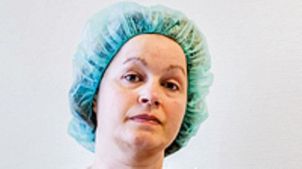
Tanja Dicht, Left Knee Patient
Dr Matthias Buhs, Orthopaedic Surgeon near Hamburg, suggested that Tanja’s damaged knee cartilage be restored via a 2-step process involving knee cartilage autotransplantation.
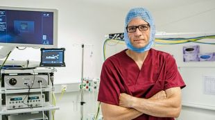
Tanja’s Orthopaedic Surgeon, Dr Matthias Buhs
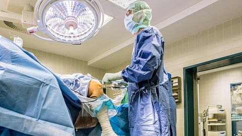
Tanja’s Autologous Knee Cartilage Transplantation and Repair
In July 2017, Tanja underwent minimally-invasive keyhole surgery called arthroscopy. This involved a small incision on each side of her kneecap.
Two small samples of healthy cartilage no bigger than grains of rice and some underlying bone were removed from the upper, non-weight-bearing parts of Tanja’s knee joint (i.e. from her distal femur).
Nicknamed white gold, these cartilage samples plus 200ml blood were sent to a laboratory in Berlin called CO.DON.** This laboratory specialises in the cultivation of hyaline articular cartilage cells.
After isolation in the laboratory, Tanja’s cartilage cells were placed via pipette into a bottle that contains a cell culture. Tanja’s bottle was then placed inside an incubator at an ambient temperature of 37°C.
There are 84 incubators in total, enabling bottles for up to 84 patients to be incubated.
Andreas Eberle of CO.DON simplified the cell incubation process as follows (translated):
‘Just imagine an oven … First, you open the door. You place the bottles inside. Then you close the door.’
During incubation, Tanja’s cartilage cells were soaked with her own donated blood serum every 2 days.
Once the bottom of the bottle was covered with newly cultivated cells, they were harvested and transferred onto microplates that contain 96 small ‘wells’ or depressions.***
A ball comprising ~200,000 chondrocytes will grow inside each well after 5 to 8 weeks of incubation. These balls are sometimes called chondrospheres, chondrocyte spheroids and/or spherical aggregates of chondrocytes (or ‘Spherox’).
Following maturation, Tanja’s microplate including 96 chondrospheres was sent to Dr Matthias Buhs for transplantation.
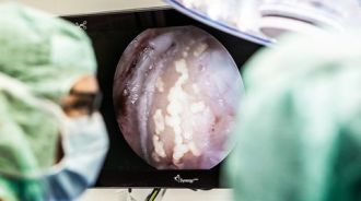
A monitor displaying a ball of cartilage cells (chondrosphere), post-transplantation
Exactly 58 days after removal of healthy cartilage samples, Tanja returned for her second minimally-invasive surgery involving autologous cartilage (chondrosphere) transplantation.
First, Tanja’s defective cartilage had to be cleaned to the bone to make way for the newly cultivated chondrospheres.
Thereafter, each ball of chondrocytes was gently transplanted into the gap. Care was taken to ensure that ‘chondrosphere overcrowding’ did not occur.
Dr Buhs watched the repaired tissue area for 20 minutes to ensure that the transplanted cell balls grew into the gap.
After the chondrospheres ‘rose like yeast dumplings’, Dr Buhs closed the incisions in Tanja’s left knee.
Thereafter, Tanja remained in bed for 3 days to enable the new chondrospheres to integrate into the native cartilage.
Summary
While non-weight-bearing gentle movements were allowed, Tanja had to avoid bearing weight on her left knee for 6 weeks.
After 6 weeks, Tanja was allowed to bear normal weight on her left knee for 6 months. Thereafter, she was able to go on walks and pursue light activities.
In Dr Buhs’ opinion, there was a 90% to 95% chance that Tanja’s left knee would be fully healed after 9 to 12 months.
On 27 March 2019, Dr Matthias Buhs confirmed that Tanja’s left knee was 100% pain free and functional.
For more details, see:
Transplantation von Knorpelgewebe – Weißes Gold: Warum Knorpel im Knie so kostbar sind. [Translated: Cartilage Tissue Transplantation – White Gold: Why knee cartilage is so precious.] (in German).
PATIENT 2 – STEPHANIE OSTERLOH
Overview
As an operations nurse, Stephanie is constantly on her feet. However, her knee was painful and swollen with limited mobility.
Stephanie first started having knee problems when she was 15 after playing handball almost daily.
Prof Dr Karl-Dieter Heller, Orthopaedic Surgeon in Braunschweig, confirmed that her cartilage was damaged and that there was too much friction.
Given the size of Stephanie’s defective cartilage site, Dr Heller recommended that she undergo autologous cartilage transplantation.
Stephanie’s Autologous Cartilage Transplantation and Repair
The first step involved removal of the damaged cartilage pieces, being the source of her knee pain. The underlying bone remained untouched.
The next step involved the extraction of a small piece of healthy cartilage and its underlying bone. This sample was sent to a laboratory for the cultivation of new cartilage cells.
The tissue sample was cut into little pieces and placed into a nutrient solution by laboratory technicians. New cartilage cells grew onto a specialised chondrocyte matrix within 3 to 6 weeks.
The cultivated cartilage cells were sent to Dr Heller who then transplanted them into Stephanie’s defective cartilage site. Once the defective site was filled, the newly cultivated cartilage cells grew into its surroundings.
Summary
According to Dr Heller (quoting; translated):
‘This treatment can only work:
- If the defect is not too large; and
- If the surrounding cartilage is completely healthy.
Otherwise it makes no sense.’
Good news! Stephanie’s treatment was a success! Her knee feels good and she can finally return to work without restriction.
After one year, Stephanie’s transplanted cartilage cells will (likely) offer the same weight-bearing capacity as her original cartilage.
For more details, see:
Walker, Niels. Knorpel defekt: Wann hilft Transplantation? [Translated: Cartilage defect: When does transplantation help?] NDR.de (10.12.2018) (in German).
https://www.ndr.de/ratgeber/gesundheit/Transplantation-bei-Knorpelschaden-im-Knie,knorpel104.html
(Includes links to two 5-minute YouTube videos, both in German)
AUTOLOGOUS OSTEOCHONDRAL TRANSFER
ELKE GREIS
Elke Greis, an artist, suffered limited mobility due to knee pain including stabbing pain.
Given that Elke injured her knee during an accident when she was 16, she was worried that 41 years later, she would need a total knee replacement.
Prof Dr Jürgen Bruns of Hamburg put Elke’s mind at ease by saying that her knee pain was due to damaged cartilage. Part of her bone was completely devoid of cartilage while another part resembled a ‘frayed, well-worn floor mat’ [translated]. Furthermore, Elke’s cartilage was damaged in a weight-bearing area. If left untreated, this could lead to arthritis.
Dr Bruns recommended that her damaged cartilage be repaired via autologous osteochondral transfer.
Lasting around 3 hours, the operation replaced Elke’s damaged cartilage with autologous (i.e. her own) healthy osteochondral tissue.
The 1st step involved removing all of Elke’s damaged cartilage.
Thereafter, a slightly larger cylindrical piece of cartilage and its underlying bone was taken from a healthy, non-weight-bearing part of Elke’s knee. The resulting hole was filled with artificial tissue while the healthy piece was seamlessly pressed into place in the defective area.
According to Dr Bruns, one cannot build a door without a door frame. In other words, one cannot transfer healthy cartilage without its underlying bone.
For optimal results, the following criteria must be met:
- There must be adequate healthy cartilage available; and
- This cartilage must match the characteristics and form of the intact cartilage near the defect area.
This is difficult to do in a joint with many curves, as in a knee.
In one year, Elke’s transferred cartilage had settled in nicely inside the defective site. Elke was now able to concentrate on her art for 1 or 2 hours at a time. Thereafter, Elke was able to walk effortlessly down the stairs. She was no longer limited by knee pain.
For more details, see:
Walker, Niels. Knorpel defekt: Wann hilft Transplantation? [Translated: Cartilage defect: When does transplantation help?] NDR.de (10.12.2018) (in German).
https://www.ndr.de/ratgeber/gesundheit/Transplantation-bei-Knorpelschaden-im-Knie,knorpel104.html
(Includes links to two 5-minute YouTube videos, both in German)
ARTICULAR CARTILAGE STEM CELL PASTE GRAFT AND THE ‘BIOKNEE’ (DEVELOPED BY DR STONE)
A 2- TO 12-YEAR FOLLOW-UP OF PASTE GRAFTING TO DEFECTIVE KNEE CARTILAGE
Dr Stone and his team followed up 125 knee patients(136 procedures; 34% female, 66% male; aged between 17 to 73) who were treated with an articular cartilage paste graft for Outerbridge grade IV arthritic chondral defects+. The authors reported that 85% of 125 knee patients enjoyed 1 grade of improvement or better following paste graft treatment.
Positive results were also observed in regenerated cartilage biopsies done via second-look arthroscopy in 66 patients. Specifically, the articular surfaces had regenerated in 42 of 66 (63.6%) biopsies. Furthermore, there was development of cartilage in 18 of 66 (27.3%) biopsies.
The authors concluded that paste grafting via minimally-invasive surgery may be a cost-effective arthroscopic treatment for patients with chronic Outerbridge classification grade IV chondral lesions.
Thus, patients with painful chondral defects in arthritic and/or traumatically-injured knees may benefit from paste graft treatment that may offer lasting pain relief, restored function and potential cartilage tissue regeneration (Stone et al, 2006).
THE ‘BIOKNEE’ (DEVELOPED BY DR STONE)
The paste graft technique is the cornerstone of Dr Stone’s ‘BioKnee’ (a possible alternative to total knee replacement).
Specifically, the ‘BioKnee’ was developed by Dr Stone for patients who have an arthritic knee that no longer has load-bearing cartilage in the knee joint nor meniscus@ tissue.
The ‘BioKnee’ involves:
- An articular cartilage paste graft; plus
- A meniscus transplantation (based on donor meniscus tissue).
More details about the ‘Bioknee’ are available here:
OTHER INTERESTING POINTS
AUTOLOGOUS CARTILAGE SOURCED ELSEWHERE
Sometimes cartilage tissue is harvested for cultivation from other autologous sources such as the nose or a rib. A Swiss study involving 20 patients obtained autologous cartilage tissue from the nose. The nasal chondrocytes were isolated, cultivated and subsequently transplanted to repair defective knee cartilage in an otherwise healthy knee joint (Caluori, University of Basel). Prof. Dr Karl-Heinz Frosch once sourced cartilage from a patient’s rib to repair defective cartilage in the same patient’s knee joint.
MESENCHYMAL STEM CELLS (MSC’s)
The progenitor of chondrocytes, being MSC’s, originate and reside in peri-articular bone marrow and subchondral bone. Mesenchymal stem cells are also present in cartilage, synovium, synovial fluid, infrapatellar/sub-synovial fat pad and adipose tissue (Mastbergen, 2013; McGonagle et al, 2017).
Recent research shows that stable cartilage tissue may also be generated from adult MSC’s following manipulation (University of Basel, 2018; University of Basel, 2018, in German).
A question:
Could adult MSC’s be a source of cultivated chondrocytes (in addition to, or instead of, healthy cartilage)?
SUMMARY
Tanja, Stephanie and Elke had positive experiences following autologous chondrocyte transplantation or autologous osteochondral transfer. These minimally-invasive options were possible because their damaged cartilage tissues were small. Therefore, their cartilage defects were repaired before severe arthritis set in.
Furthermore, 85% of 125 knee patients enjoyed 1 grade of improvement or better following articular cartilage stem cell paste graft treatment by Dr Stone at The Stone Clinic in San Francisco.
Finally, using autologous (i.e. one’s own) cartilage tissue ensures nil risk of rejection by the immune system.
I hope that these patient stories offer hope to people with knee pain due to defective cartilage in an otherwise healthy knee joint.
Sabina Walker
Blogger, Pain Matters (in WordPress)
Please forward to anyone who may benefit from this information.
KEY
* Autologous cartilage cell transplantation (aka Autologous chondrocyte transplantation) = Where the patient uses his/her own knee cartilage for propagation ex vivo and transplantation.
NB Cartilage cells are also called chondrocytes.
** CO.DON specialises in the cultivation of autologous cartilage cells for minimally-invasive repair of defects in knee cartilage tissue.
http://www.codon.de/home.html?L=1
Other tissue engineering companies including Tetec also offer this service.
https://www.tetec-ag.com/en.html
*** Microplates, also called microtiter plates, are flat plates with many small ‘wells’ (depressions, or hollows).
**** Chondrocytes are the cartilage-producing cells in the body (more simply, cartilage cells).
# Microfracture surgery involves making small holes in the bone(s) of a knee joint to create bleeding and release marrow cells into defective cartilage. This may induce a healing response and the growth of new cartilage.
@ The meniscus is C-shaped cartilage tissue that acts as a shock absorber between the shin bone (tibia) and thigh bone (femur).
$ The intercondylar notch of femur is a depression between the posterior surfaces of the medial and lateral epicondyle of the thigh bone at the knee joint.
+ Outerbridge grade IV chondral defects are cartilage lesions (or erosions) right to the underlying subchondral bone.
REFERENCES
Media
(1) Buhs, Matthias. Cartilage Center of Northern Germany
https://www.cartilagecenter.de/dr-matthias-buhs/
(2A) NICE. Autologous chondrocyte implantation using chondrosphere for treating symptomatic articular cartilage defects of the knee. NICE (7 March 2018).
https://www.nice.org.uk/guidance/TA508/chapter/1-Recommendations
(2B) Spherox – Spheroids of human autologous matrix-associated chondrocytes. European Medicines Agency
https://www.ema.europa.eu/en/medicines/human/EPAR/spherox
(3A) Washington University Orthopedics. Autologous Chondrocyte Implantation (ACI). Washington University.
(3B) Keeffe, Patrick. Knee Surgeries Could Soon Be More Successful Thanks to New Technique. Healthline (14 October 2018).
(4A) Caluori, Reto. From nose to knee. University of Basel.
https://www.unibas.ch/en/Research/Uni-Nova/Uni-Nova-129/Uni-Nova-129-From-nose-to-knee.html
(4B) BIOengineered grafts for Cartilage Healing In Patients (BIO-CHIP)
(5A) Stone, KR. Study proves stem cell paste graft therapy critical to rebuilding cartilage. The Stone Clinic.
https://www.stoneclinic.com/stem-cell-cartilage-study
(Above link includes 2 short videos called Articular Cartilage Paste Graft and Articular Cartilage Paste Graft Surgical Technique.)
(5B) Stone, KR. Why Microfracture Fails. The Stone Clinic (7 Feb 2015).
https://www.stoneclinic.com/blog/why-microfracture-fails
Media (in German)
(1) Schneider, Mathias. Transplantation von Knorpelgewebe – Weißes Gold: Warum Knorpel im Knie so kostbar sind. [Translated: Cartilage Tissue Transplantation – White Gold: Why knee cartilage is so precious.] Der Stern (7 October 2017); 80-84.
http://mobil.stern.de/gesundheit/medizin/knie–knorpel-im-knie-sind-so-kostbar-wie-kaum-ein-anderes-gewebe-7645258.html
(2) Walker, Niels. Knorpel defekt: Wann hilft Transplantation? [Translated: Cartilage defect: When does transplantation help?] NDR.de (10.12.2018) (in German).
https://www.ndr.de/ratgeber/gesundheit/Transplantation-bei-Knorpelschaden-im-Knie,knorpel104.html
(Includes links to two 5-minute YouTube videos, both in German)
(3) University of Basel. Züchtung von Knorpel aus Stammzellen gelungen. aerzteblatt.de (18/4/18).
https://www.aerzteblatt.de/nachrichten/93534/Zuechtung-von-Knorpel-aus-Stammzellen-gelungen
Peer-Reviewed Papers
(1) Piñeiro-Ramil et al. Cell Therapy and Tissue Engineering for Cartilage Repair. IntechOpen (20 Dec 2017).
doi: 10.5772/intechopen.70406
(2A) Developmentally inspired programming of adult human mesenchymal stromal cells toward stable chondrogenesis.
(2B) University of Basel. Cultivating cartilage from stem cells. Science Daily (16 April 2018).
https://www.sciencedaily.com/releases/2018/04/180416155622.htm
(3) Peterson et al. Two- to 9-year outcome after autologous chondrocyte transplantation of the knee. Clin Orthop Relat Res (May 2000); 374: 212-34.
(4A) Huey D., Hu J., Athanasiou K. (2012) Unlike bone, cartilage regeneration remains elusive. Science338: 917–921.
https://www.ncbi.nlm.nih.gov/pmc/articles/PMC4327988/
(4B) Tiku ML, Sabaawy HE. Cartilage regeneration for treatment of osteoarthritis: a paradigm for nonsurgical intervention. Ther Adv Musculoskelet Dis. 2015;7(3):76-87.
https://www.ncbi.nlm.nih.gov/pmc/articles/PMC4426098/
(4C) S.R. Lyu, D.S. Liu, C.E. Tseng, H.S. Wang and L.K. Chau (2012). Knee Health Promotion Option for Osteoarthritic Knee: Cartilage Regeneration is Possible, Osteoarthritis – Diagnosis, Treatment and Surgery, Prof. Qian Chen (Ed.), ISBN: 978-953-51-0168-0, InTech, Available from: http://www.intechopen.com/books/osteoarthritis-diagnosis-treatment-and-surgery/knee-health-promotion- option-for-osteoarthritic-knee-cartilage-regeneration-is-possible
(5) Brittberg et al. Matrix-Applied Characterized Autologous Cultured Chondrocytes Versus Microfracture: Five-Year Follow-up of a Prospective Randomized Trial. The American Journal of Sports Medicine (1 May 2018); 46(6): 1343-1351.
(6A) Stone, KR et al. Articular Cartilage Paste Grafting to Full-Thickness Articular Cartilage Knee Joint Lesions: A 2- to 12-Year Follow-up. Arthroscopy: The Journal of Arthroscopic and Related Surgery (March 2006); 22(3): 291-299.
https://www.stoneclinic.com/sites/default/files/uploads/stone.arthroscopy2006.artcart2-12yr.pdf
(6B) Xing, L et al. Microfracture combined with osteochondral paste implantation was more effective than microfracture alone for full-thickness cartilage repair. Knee Surg Sports Traumatol Arthrosc (Aug 2013); 21(8): 1770-76.
https://doi.org/10.1007/s00167-012-2031-5
(7A) Mastbergen, S. SP0166 Joint Distraction and Cartilage Regeneration – What is the Basis for Structural Repair? Ann Rheum Dis (2013); 72(Suppl 3).
http://dx.doi.org/10.1136/annrheumdis-2013-eular.166
https://ard.bmj.com/content/72/Suppl_3/A38.2
(7B) McGonagle et al. Native joint-resident mesenchymal stem cells for cartilage repair in osteoarthritis. Nat Rev Rheumatol (Dec 2017); 13(12): 719-730.
doi 10.1038/nrrheum.2017.182.
